

Isolation of a phosphonate-degrading symbiotic bacterium from Prorocentrum donghaiense and its promoting effect on algal growth
-
摘要: 海洋中的膦酸酯(C-P键有机磷)是可供浮游植物利用的一种潜在磷源。甲藻自身无法直接利用膦酸酯,但是其共生细菌可将膦酸酯降解为磷酸盐,从而促进藻细胞的生长。然而,目前尚未有针对特定菌株的相关研究。本研究在2-氨基乙基膦酸(2-AEP)作为唯一磷源条件下对东海原甲藻进行培养,对其中的共生细菌进行分离纯化,初步得到5种细菌。基因组测序结果表明,其中一株尹氏菌属细菌Yoonia sp. PD-AEP-1中存在两种C-P裂解酶途径。通过藻菌共培养实验对菌株功能进行验证,结果显示,将藻细胞处理至磷饥饿状态之后,同时加入2-AEP与PD-AEP-1悬液,与只加2-AEP或只加细菌悬液的条件相比,藻细胞生长速率和体系内磷酸盐浓度显著升高,同时碱性磷酸酶活性及非光化学淬灭值则明显降低,表明PD-AEP-1具备将2-AEP降解为磷酸盐的能力,进而缓解了东海原甲藻细胞的磷限制状态,有效促进了藻细胞的生长。该研究表明,东海原甲藻共生细菌在降解膦酸酯从而为藻细胞提供磷源方面扮演着一定角色,这一过程很可能有助于东海原甲藻赤潮的暴发,凸显了海洋生态系统中藻−菌相互作用的重要性。Abstract: Phosphonates in the ocean are a kind of potential phosphorus (P) source which could be utilized by phytoplankton. Although dinoflagellates cannot directly utilize phosphonates themselves, their symbiotic bacteria have the capability to degrade phosphonates into phosphate, thereby promoting the growth of algal cells. However, no studies focusing on a specific bacteria strain have been conducted thus far. In this study, Prorocentrum donghaiense was cultured under conditions with 2-Aminoethylphosphonic acid (2-AEP) as the sole P source. Isolation and purification of the symbiotic bacteria from the culture was conducted and five kinds of bacteria were obtained. Genome sequencing results revealed the presence of two types of C-P lyase pathways in the bacterial strain designated as Yoonia sp. PD-AEP-1. The function of the bacteira strain was verified through the co-culture of bacteria and algal cells. The results demonstrated that after the algal cells were treated to phosphorus-starved condition, when 2-AEP and the bacteria suspension were added together, as compared to conditions which only 2-AEP or the bacterial suspension of PD-AEP-1 was introduced, both the growth rate of algal cells and the phosphate concentration in the cultures showed a significant increase. Meanwhile, alkaline phosphatase activity and non-photochemical quenching of the algal cells decreased significantly, indicating that PD-AEP-1 has the ability to degrade 2-AEP into phosphate, thereby alleviating phosphorus limitation for P. donghaiense cells and effectively promoting the growth of algal cells. The study suggests that symbiotic bacteria of P. donghaiense might play a part in providing P sources to the algal cells through the degradation of phosphonates. This process could probably contribute to the outbreak of P. donghaiense bloom, highlighting the importance of algae-bacteira interactions in marine ecosystems.
-
Key words:
- phosphonates /
- Prorocentrum donghaiens /
- symbiotic bacteria /
- algal-growth-promoting effect /
- red tide
-

图 1 3种磷源条件下东海原甲藻细胞的生长曲线(a)及培养体系内磷酸盐浓度(b)
磷酸盐:f/2培养基,磷酸盐浓度为36 μmol/L;2-AEP:f/2-P培养基基础上添加36 μmol/L的2-AEP;无磷:f/2-P培养基,未添加任何磷源
Fig. 1 Growth curve of Prorocentrum donghaiense (a) and phosphate concentration in the culture (b) under three P conditions
Phosphate: f/2 medium, phosphate concentration was 36 μmol/L; 2-AEP: f/2-P medium, while 36 μmol/L 2-AEP was added; No phosphorus: f/2-P medium, no P source was added
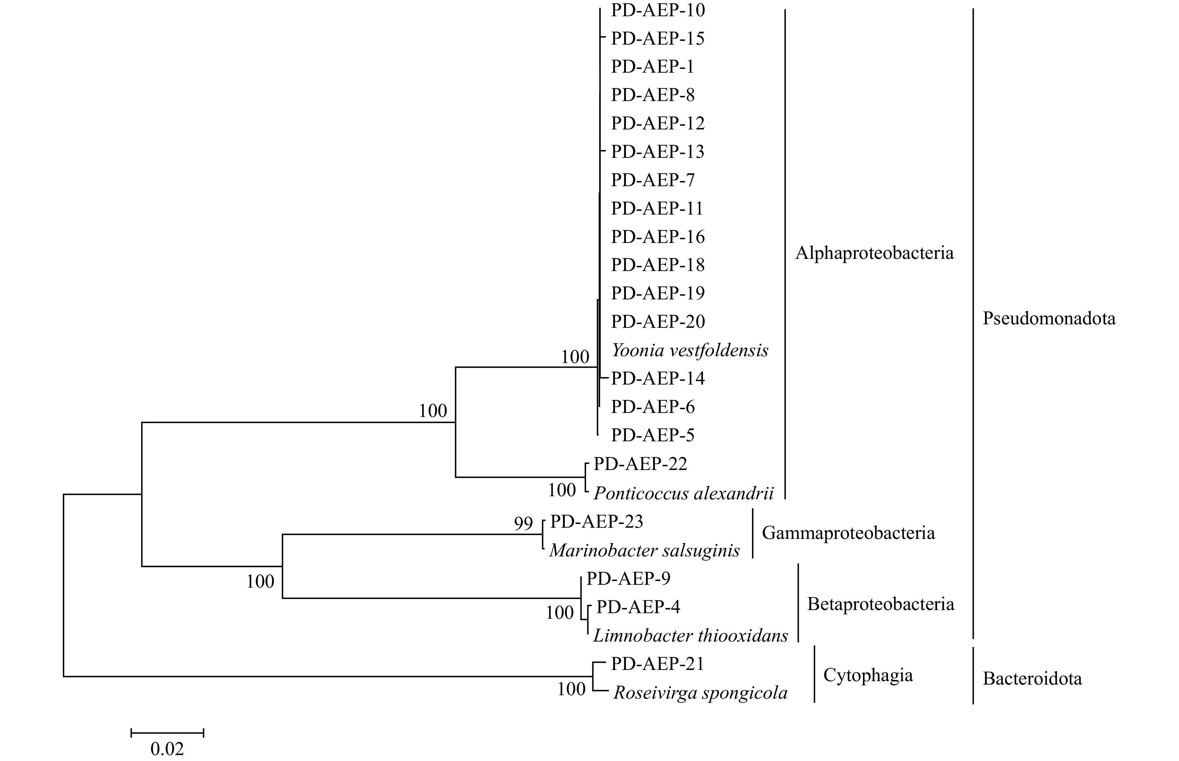
图 2 基于16S rRNA基因序列构建的东海原甲藻共生细菌及其相近菌株Neighbour-Joining树
Fig. 2 Neighbour-Joining tree of Prorocentrum donghaiense symbiotic bacteria and their close strains constructed in the basis of the 16S rRNA gene sequences

图 3 PD-AEP-1基因组中归属于RAST不同子系统类别基因的分布情况
Fig. 3 Distribution of the genes from the genome of PD-AEP-1 which belong to the “Subsystem Category” of RAST

图 4 PD-APE-1基因组中两个C-P裂解酶途径基因簇的排布
“*”表示基因簇II中的基因;不同颜色分别代表编码膦酸酯转运蛋白(绿色)、调控蛋白(紫色)及C-P裂解酶亚基蛋白的基因,黑色为未知功能蛋白;atf: 膦酸酯利用相关乙酰转移酶基因, atu: 未知功能蛋白Atu0170编码基因
Fig. 4 The organization of two gene clusters of C-P lyase pathways in the genome of the PD-AEP-1
“*” indicates the genes of the second cluster. Genes encoding phosphonate transport (green) and regulation (purple), the C-P lyase subunits (yellow) and other proteins of unknown function (black) are shown in different colors. atf: phosphonate utilization associated acetyltransferase, atu: uncharacterized protein Atu0170
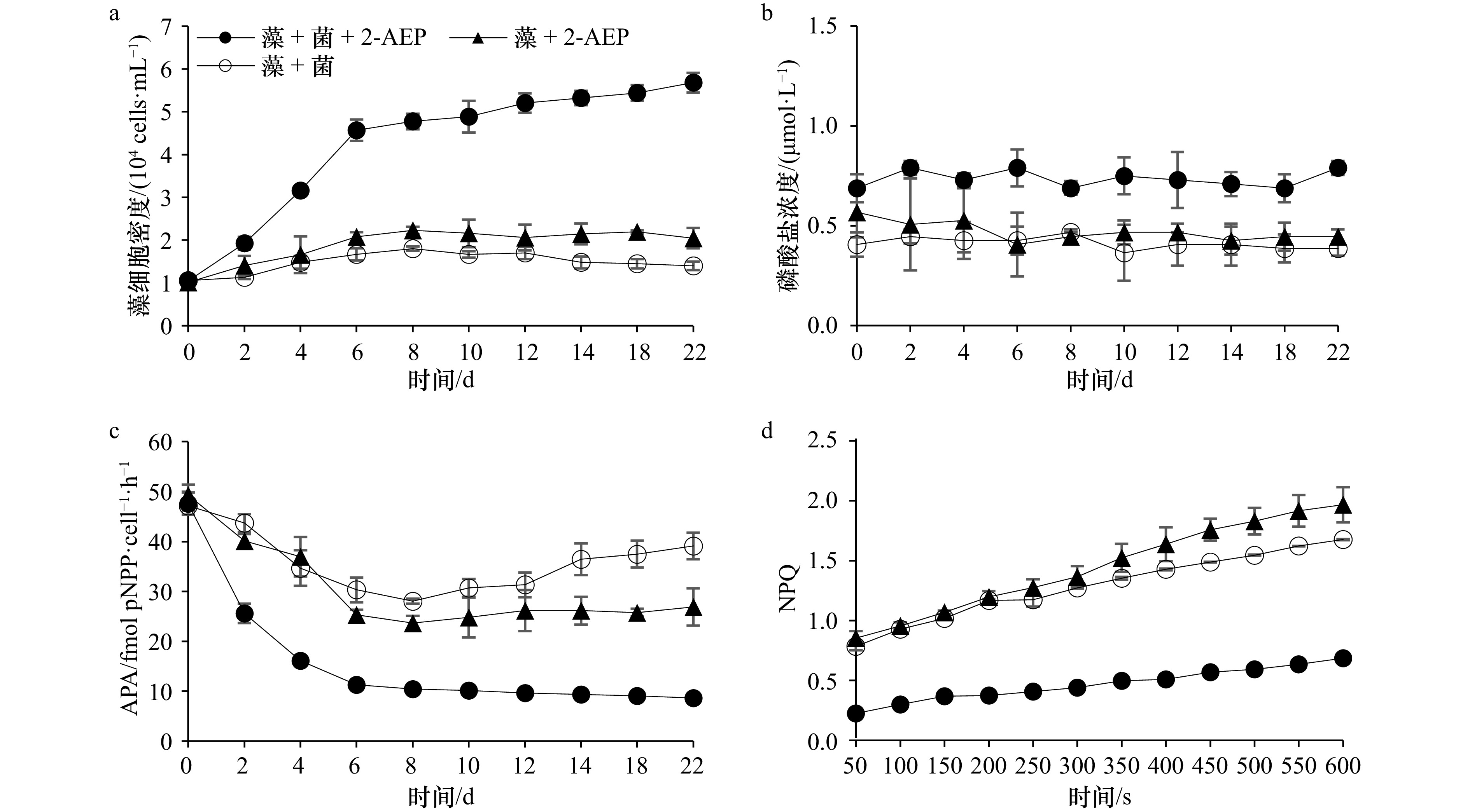
图 5 3种培养条件下东海原甲藻细胞的生长曲线(a)、磷酸盐浓度(b)、碱性磷酸酶活性(c)及第12 d的NPQ诱导曲线(d)
藻 + 菌:藻细胞培养体系中加入1%体积细菌悬液;藻 + 2-AEP:藻细胞培养体系中加入36 μmol/L 2-AEP;藻 + 菌 + 2-AEP:藻细胞培养体系中同时加入1%体积细菌悬液和36 μmol/L 2-AEP
Fig. 5 Growth curve and of Prorocentrum donghaiense, phosphate concentration measurement, alkaline phosphatase activity (APA) measurement and induction curve of NPQ in the 12th day under three culture conditions
Algae + bacteria: algal culture systems added with 1% volume of bacteria suspension; Algae + 2-AEP: algal culture systems added with 36 μmol/L 2-AEP; Algae + bacteria + 2-AEP: algal culture systems added with 1% volume of bacteria suspension and 36 μmol/L 2-AEP
表 1 PD-AEP-1基因组中与磷代谢相关的基因及其特征
Tab. 1 Genes and their characteristics related to P metabolism in the genome of PD-AEP-1
子系统 基因编号 功能 高亲和力磷酸盐转运蛋白和 Pho 调节子调控 peg.1464 磷酸盐转运系统调节蛋白PhoU peg.1463 磷酸盐调节子转录调节蛋白PhoB (SphR) peg.1832 磷酸盐调节子传感蛋白PhoR (SphS) (EC 2.7.13.3) 多聚磷酸盐 peg.2944 聚磷酸盐激酶 (EC 2.7.4.1) peg.2027 外切聚磷酸酶 (EC 3.6.1.11) 磷酸盐代谢 peg.1508 NAD(P) 转氢酶β亚基 (EC 1.6.1.2) peg.3897 碱性磷酸酶 (EC 3.1.3.1) peg.3045 磷酸盐饥饿诱导蛋白PhoH peg.1039 锰依赖型无机焦磷酸酶 (EC 3.6.1.1)  下载: 导出CSV
表 2 PD-AEP-1基因组中与膦酸酯利用相关的基因及其特征
Tab. 2 Genes related to phosphonate utilization in the genome of PD-AEP-1 and their characteristics
重叠群 基因编号 长度/bp 编码的酶/蛋白* NODE_9_length_69264_cov_65.868739 peg.3912 1146 Alpha-D-ribose 1-methylphosphonate 5-triphosphate diphosphatase PhnM2 (EC 3.6.1.63) peg.3913 675 Uncharacterized protein Atu0170, clustered with phosphonate utilization peg.3914 543 Ribose 1,5-bisphosphate phosphokinase PhnN (EC 2.7.4.23) peg.3915 684 Alpha-D-ribose 1-methylphosphonate 5-triphosphate synthase subunit PhnL (EC 2.7.8.37) peg.3916 771 Phosphonates utilization ATP-binding protein PhnK peg.3917 831 Alpha-D-ribose 1-methylphosphonate 5-phosphate C-P lyase PhnJ (EC 4.7.1.1) peg.3918 1083 Alpha-D-ribose 1-methylphosphonate 5-triphosphate synthase subunit PhnI (EC 2.7.8.37) peg.3919 561 Alpha-D-ribose 1-methylphosphonate 5-triphosphate synthase subunit PhnH (EC 2.7.8.37) peg.3920 456 Alpha-D-ribose 1-methylphosphonate 5-triphosphate synthase subunit PhnG (EC 2.7.8.37) peg.3921 723 Transcriptional regulator PhnF peg.3922 1188 Metal-dependent hydrolase involved in phosphonate metabolism PhnM1 peg.3923 615 Phosphonate utilization associated acetyltransferase (ATF) peg.3924 1329 Phosphonate ABC transporter permease protein PhnE1 (TC 3.A.1.9.1) peg.3925 873 Phosphonate ABC transporter permease protein PhnE2 (TC 3.A.1.9.1) peg.3926 903 Phosphonate ABC transporter substrate-binding protein PhnD (TC 3.A.1.9.1) peg.3927 819 Phosphonate ABC transporter ATP-binding protein PhnC (TC 3.A.1.9.1) NODE_1_length_861959_cov_71.815100 peg.775 537 PhnH protein peg.776 1056 Alpha-D-ribose 1-methylphosphonate 5-triphosphate synthase subunit PhnI (EC 2.7.8.37) peg.777 915 Alpha-D-ribose 1-methylphosphonate 5-phosphate C-P lyase PhnJ (EC 4.7.1.1) peg.778 798 Phosphonates utilization ATP-binding protein PhnK peg.779 720 Alpha-D-ribose 1-methylphosphonate 5-triphosphate synthase subunit PhnL (EC 2.7.8.37) peg.780 1194 Alpha-D-ribose 1-methylphosphonate 5-triphosphate diphosphatase PhnM (EC 3.6.1.63) peg.781 816 Phosphonate ABC transporter ATP-binding protein PhnC (TC 3.A.1.9.1) peg.782 915 Phosphonate ABC transporter substrate-binding protein PhnD (TC 3.A.1.9.1) peg.783 933 ABC transporter, permease protein PhnE1 peg.784 858 Phosphonate ABC transporter permease protein PhnE2(TC 3.A.1.9.1) peg.785 630 Phosphonate utilization associated acetyltransferase NODE_24_length_14478_cov_37.311755 peg.1318 576 PhnH protein peg.1319 1014 Alpha-D-ribose 1-methylphosphonate 5-triphosphate synthase subunit PhnI (EC 2.7.8.37) peg.1320 846 Alpha-D-ribose 1-methylphosphonate 5-phosphate C-P lyase PhnJ (EC 4.7.1.1) peg.1321 768 Phosphonates utilization ATP-binding protein PhnK 注:因该列酶和蛋白的名称仍然未有公认的中文译名,因此采用英文名称。
下载: 导出CSV
-
[1] Tyrrell T. The relative influences of nitrogen and phosphorus on oceanic primary production[J]. Nature, 1999, 400(6744): 525−531. doi: 10.1038/22941 [2] Karl D M. Microbially mediated transformations of phosphorus in the sea: new views of an old cycle[J]. Annual Review of Marine Science, 2014, 6: 279−337. doi: 10.1146/annurev-marine-010213-135046 [3] Lin Senjie, Litaker R W, Sunda W G. Phosphorus physiological ecology and molecular mechanisms in marine phytoplankton[J]. Journal of Phycology, 2016, 52(1): 10−36. doi: 10.1111/jpy.12365 [4] Kolowith L C, Ingall E D, Benner R. Composition and cycling of marine organic phosphorus[J]. Limnology and Oceanography, 2001, 46(2): 309−320. doi: 10.4319/lo.2001.46.2.0309 [5] Lin Xin, Wang Lu, Shi Xinguo, et al. Rapidly diverging evolution of an atypical alkaline phosphatase (PhoA(aty)) in marine phytoplankton: insights from dinoflagellate alkaline phosphatases[J]. Frontiers in Microbiology, 2015, 6: 868. [6] McGrath J W, Chin J P, Quinn J P. Organophosphonates revealed: new insights into the microbial metabolism of ancient molecules[J]. Nature Reviews Microbiology, 2013, 11(6): 412−419. doi: 10.1038/nrmicro3011 [7] Gomez-Garcia M R, Davison M, Blain-Hartnung M, et al. Alternative pathways for phosphonate metabolism in thermophilic cyanobacteria from microbial mats[J]. The ISME Journal, 2011, 5(1): 141−149. doi: 10.1038/ismej.2010.96 [8] Dyhrman S T, Chappell P D, Haley S T, et al. Phosphonate utilization by the globally important marine diazotroph Trichodesmium[J]. Nature, 2006, 439(7072): 68−71. doi: 10.1038/nature04203 [9] Whitney L P, Lomas M W. Phosphonate utilization by eukaryotic phytoplankton[J]. Limnology and Oceanography Letters, 2019, 4(1): 18−24. doi: 10.1002/lol2.10100 [10] Wang Cong, Lin Xin, Li Ling, et al. Differential growth responses of marine phytoplankton to herbicide glyphosate[J]. PLoS One, 2016, 11(3): e0151633. doi: 10.1371/journal.pone.0151633 [11] Cui Yudong, Lin Xin, Zhang Huan, et al. PhnW-PhnX pathway in dinoflagellates not functional to utilize extracellular phosphonates[J]. Frontiers in Marine Science, 2016, 2: 120. [12] Yu Xiaomin, Doroghazi J R, Janga S C, et al. Diversity and abundance of phosphonate biosynthetic genes in nature[J]. Proceedings of the National Academy of Sciences of the United States of America, 2013, 110(51): 20759−20764. [13] Dyhrman S T, Benitez-Nelson C R, Orchard E D, et al. A microbial source of phosphonates in oligotrophic marine systems[J]. Nature Geoscience, 2009, 2(10): 696−699. doi: 10.1038/ngeo639 [14] Quin L D, Quin G S. Screening for carbon-bound phosphorus in marine animals by high-resolution 31P-NMR spectroscopy: coastal and hydrothermal vent invertebrates[J]. Comparative Biochemistry and Physiology Part B: Biochemistry and Molecular Biology, 2001, 128(1): 173−185. doi: 10.1016/S1096-4959(00)00310-9 [15] Wang Cong, Lin Xin, Li Ling, et al. Glyphosate shapes a dinoflagellate-associated bacterial community while supporting algal growth as sole phosphorus source[J]. Frontiers in Microbiology, 2017, 8: 2530. doi: 10.3389/fmicb.2017.02530 [16] Quinn J P, Kulakova A N, Cooley N A, et al. New ways to break an old bond: the bacterial carbon–phosphorus hydrolases and their role in biogeochemical phosphorus cycling[J]. Environmental Microbiology, 2007, 9(10): 2392−2400. doi: 10.1111/j.1462-2920.2007.01397.x [17] Villarreal-Chiu JF, Quinn JP, McGrath JW. The genes and enzymes of phosphonate metabolism by bacteria, and their distribution in the marine environment[J]. Frontiers in Microbiology, 2012, 3: 19. [18] Richardson B, Corcoran A A. Use of dissolved inorganic and organic phosphorus by axenic and nonaxenic clones of Karenia brevis and Karenia mikimotoi[J]. Harmful Algae, 2015, 48: 30−36. doi: 10.1016/j.hal.2015.06.005 [19] Shi Xinguo, Lin Xin, Li Ling, et al. Transcriptomic and microRNAomic profiling reveals multi-faceted mechanisms to cope with phosphate stress in a dinoflagellate[J]. The ISME Journal, 2017, 11(10): 2209−2218. doi: 10.1038/ismej.2017.81 [20] Shi Xinguo, Liu Lenian, Li Yue, et al. Isolation of an algicidal bacterium and its effects against the harmful-algal- bloom dinoflagellate Prorocentrum donghaiense (Dinophyceae)[J]. Harmful Algae, 2018, 80: 72−79. doi: 10.1016/j.hal.2018.09.003 [21] Parkhill J P, Maillet G, Cullen J J. Fluorescence-based maximal quantum yield for PSII as a diagnostic of nutrient stress[J]. Journal of Phycology, 2001, 37(4): 517−529. doi: 10.1046/j.1529-8817.2001.037004517.x [22] Baker N R. Chlorophyll fluorescence: a probe of photosynthesis in vivo[J]. Annual Review of Plant Biology, 2008, 59: 89−113. doi: 10.1146/annurev.arplant.59.032607.092759 [23] Tamura K, Stecher G, Peterson D, et al. MEGA6: molecular evolutionary genetics analysis version 6.0[J]. Molecular Biology and Evolution, 2013, 30(12): 2725−2729. doi: 10.1093/molbev/mst197 [24] Overbeek R, Olson R, Pusch G D, et al. The SEED and the rapid annotation of microbial genomes using subsystems technology (RAST)[J]. Nucleic Acids Research, 2014, 42(D1): D206−D214. doi: 10.1093/nar/gkt1226 [25] Chun J, Oren A, Ventosa A, et al. Proposed minimal standards for the use of genome data for the taxonomy of prokaryotes[J]. International Journal of Systematic and Evolutionary Microbiology, 2018, 68(1): 461−466. doi: 10.1099/ijsem.0.002516 [26] Kim M, Oh H S, Park S C, et al. Towards a taxonomic coherence between average nucleotide identity and 16S rRNA gene sequence similarity for species demarcation of prokaryotes[J]. International Journal of Systematic and Evolutionary Microbiology, 2014, 64(Pt 2): 346-351. [27] Cui Yudong, Zhang Huan, Lin Senjie. Enhancement of non-photochemical quenching as an adaptive strategy under phosphorus deprivation in the dinoflagellate Karlodinium veneficum[J]. Frontiers in Microbiology, 2017, 8: 404. [28] Oren A, Garrity G M. Valid publication of the names of forty-two phyla of prokaryotes[J]. International Journal of Systematic and Evolutionary Microbiology, 2021, 71(10): 005056. [29] 唐莹莹, 乔玉宝, 蒋志伟, 等. 东海产麻痹性贝毒链状亚历山大藻共附生菌群多样性研究[J]. 海洋渔业, 2018, 40(6): 720−727. doi: 10.3969/j.issn.1004-2490.2018.06.009Tang Yingying, Qiao Yubao, Jiang Zhiwei, et al. Biodiversity study of the bacterial community associated with toxic marine dinoflagellate Alexandrium catenella LZ1706[J]. Marine Fisheries, 2018, 40(6): 720−727. doi: 10.3969/j.issn.1004-2490.2018.06.009 [30] 龚诗雁, 屠燕萍, 谢志浩. 4株米氏凯伦藻(Karenia mikimotoi)藻际异养细菌的分离鉴定[J]. 海洋与湖沼, 2014, 45(5): 1099−1104. doi: 10.11693/hyhz20140600174Gong Shiyan, Tu Yanping, Xie Zhihao. Molecular identification of four strains of heterotrophic bacteria isolated from Karenia mikimotoi[J]. Oceanologia et Limnologia Sinica, 2014, 45(5): 1099−1104. doi: 10.11693/hyhz20140600174 [31] 李月月, 田晓清, 韩清华, 等. 利玛原甲藻PL11共附生菌多样性研究[J]. 海洋渔业, 2020, 42(1): 73−81. doi: 10.3969/j.issn.1004-2490.2020.01.008Li Yueyue, Tian Xiaoqing, Han Qinghua, et al. Biodiversity of symbiotic and epiphytic bacteria of Prorocentrum lima PL11[J]. Marine Fisheries, 2020, 42(1): 73−81. doi: 10.3969/j.issn.1004-2490.2020.01.008 [32] Wirth J S, Whitman W B. Phylogenomic analyses of a clade within the roseobacter group suggest taxonomic reassignments of species of the genera Aestuariivita, Citreicella, Loktanella, Nautella, Pelagibaca, Ruegeria, Thalassobius, Thiobacimonas and Tropicibacter, and the proposal of six novel genera[J]. International Journal of Systematic and Evolutionary Microbiology, 2018, 68(7): 2393−2411. doi: 10.1099/ijsem.0.002833 [33] 曹延群, 李赟, 潘克厚, 等. 三角褐指藻藻液细菌的分离鉴定及其对藻细胞生长的影响[J]. 海洋湖沼通报, 2019(1): 107−112.Cao Yanqun, Li Yun, Pan Kehou, et al. Isolation and identification of the bacteria from Phaeodactylum tricornutum and their effects on the growth of algal cells[J]. Transactions of Oceanology and Limnology, 2019(1): 107−112. [34] Johansson O N, Pinder M I M, Ohlsson F, et al. Friends with benefits: exploring the phycosphere of the marine diatom Skeletonema marinoi[J]. Frontiers in Microbiology, 2019, 10: 1828. doi: 10.3389/fmicb.2019.01828 [35] Buchan A, LeCleir G R, Gulvik C A, et al. Master recyclers: features and functions of bacteria associated with phytoplankton blooms[J]. Nature Reviews Microbiology, 2014, 12(10): 686−698. doi: 10.1038/nrmicro3326 [36] 刘兵, 李宇, 叶倩, 等. 链状亚历山大藻共附生细菌多样性[J]. 生态学杂志, 2009, 28(5): 889−894.Liu Bing, Li Yu, Ye Qian, et al. Phylogenetic diversity of bacteria associated with dinoflagellate Alexandrium catenella[J]. Chinese Journal of Ecology, 2009, 28(5): 889−894. [37] 杨小茹, 苏建强, 郑小伟, 等. 基于分子技术的1株产毒藻藻际细菌多样性分析[J]. 环境科学, 2009, 30(1): 271−279. doi: 10.3321/j.issn:0250-3301.2009.01.046Yang Xiaoru, Su Jianqiang, Zheng Xiaowei, et al. 16S rDNA clone library analysis of microbial diversity associated with the PSP-producing dinoflagellate Alexandrium tamarense[J]. Environmental Science, 2009, 30(1): 271−279. doi: 10.3321/j.issn:0250-3301.2009.01.046 [38] 王鹏斌, 戴鑫烽, 陆斗定. 米氏凯伦藻(Km02)共培养细菌群落的研究[J]. 海洋与湖沼, 2019, 50(3): 644−651. doi: 10.11693/hyhz20180700179Wang Pengbin, Dai Xinfeng, Lu Douding. Co-cultured bacterial community of Karenia mikimotoi (Km02)[J]. Oceanologia et Limnologia Sinica, 2019, 50(3): 644−651. doi: 10.11693/hyhz20180700179 [39] Wang Yuming, Zhou Panpan, Zhou Weicheng, et al. Network analysis indicates microbial assemblage differences in life stages of Cladophora[J]. Applied and Environmental Microbiology, 2023, 89(3): e0211222. doi: 10.1128/aem.02112-22 [40] Oppong-Danquah E, Blümel M, Tasdemir D. Metabolomics and microbiomics insights into differential surface fouling of three macroalgal species of Fucus (Fucales, Phaeophyceae) that co-exist in the German Baltic Sea[J]. Mar Drugs, 2023, 21(11): 595. doi: 10.3390/md21110595 [41] 李斯远, 何治江, 吕泓玥, 等. 厚壳贻贝(Mytilus coruscus)养殖海域与天然生长海域的微生物群落比较研究[J]. 海洋与湖沼, 2021, 52(1): 196−205. doi: 10.11693/hyhz20200700217Li Siyuan, He Zhijiang, Lv Hongyue, et al. Comparative study on microbial community in mussel Mytilus coruscus body and seawater of its natural and cultural sea area in Zhoushan, Zhejiang[J]. Oceanologia et Limnologia Sinica, 2021, 52(1): 196−205. doi: 10.11693/hyhz20200700217 [42] Du Zongjun, Zhang Wanyi, Xia Hongjie, et al. Isolation and diversity analysis of heterotrophic bacteria associated with sea anemones[J]. Acta Oceanologica Sinica, 2010, 29(2): 62−69. doi: 10.1007/s13131-010-0023-1 [43] Huo Lixin, Ma Anran, Liu Hong, et al. Diversity and ecological assembly process of aerobic anoxygenic phototrophic bacteria in a low irradiation area, three gorges reservoir[J]. Journal of Environmental Sciences, 2024, 143: 116−125. doi: 10.1016/j.jes.2023.08.015 [44] Feng Xiaoyuan, Xing Peng. Genomics of Yoonia sp. isolates (family Roseobacteraceae) from Lake Zhangnai on the Tibetan Plateau[J]. Microorganisms, 2023, 11(11): 2817. doi: 10.3390/microorganisms11112817 [45] Piontek J, Meeske C, Hassenrück C, et al. Organic matter availability drives the spatial variation in the community composition and activity of Antarctic marine bacterioplankton[J]. Environmental Microbiology, 2022, 24(9): 4030−4048. doi: 10.1111/1462-2920.16087 [46] Cruz-López R, Maske H. The vitamin B1 and B12 required by the marine dinoflagellate Lingulodinium polyedrum can be provided by its associated bacterial community in culture[J]. Frontiers in Microbiology, 2016, 7: 560. [47] Orchard E D, Benitez-Nelson C R, Pellechia P J, et al. Polyphosphate in Trichodesmium from the low‐phosphorus Sargasso Sea[J]. Limnology and Oceanography, 2010, 55(5): 2161−2169. doi: 10.4319/lo.2010.55.5.2161 [48] Dyhrman S T, Ammerman J W, Van Mooy B A S. Microbes and the marine phosphorus cycle[J]. Oceanography, 2007, 20(2): 110−116. doi: 10.5670/oceanog.2007.54 [49] Metcalf W W, Wanner B L. Evidence for a fourteen-gene, phnC to phnP locus for phosphonate metabolism in Escherichia coli[J]. Gene, 1993, 129(1): 27−32. doi: 10.1016/0378-1119(93)90692-V [50] Hove-Jensen B, Rosenkrantz T J, Zechel D L, et al. Accumulation of intermediates of the carbon-phosphorus lyase pathway for phosphonate degradation in phn mutants of Escherichia coli[J]. Journal of Bacteriology, 2010, 192(1): 370−374. doi: 10.1128/JB.01131-09 [51] White A K, Metcalf W W. Two C-P lyase operons in Pseudomonas stutzeri and their roles in the oxidation of phosphonates, phosphite, and hypophosphite[J]. Journal of Bacteriology, 2004, 186(14): 4730−4739. doi: 10.1128/JB.186.14.4730-4739.2004 [52] Errey J C, Blanchard J S. Functional annotation and kinetic characterization of PhnO from Salmonella enterica[J]. Biochemistry, 2006, 45(9): 3033−3039. doi: 10.1021/bi052297p [53] Amin S A, Hmelo L R, Van Tol H M, et al. Interaction and signalling between a cosmopolitan phytoplankton and associated bacteria[J]. Nature, 2015, 522(7554): 98−101. doi: 10.1038/nature14488 [54] González J M, Simó R, Massana R, et al. Bacterial community structure associated with a dimethylsulfoniopropionate-producing North Atlantic algal bloom[J]. Applied and Environmental Microbiology, 2000, 66(10): 4237−4246. doi: 10.1128/AEM.66.10.4237-4246.2000 [55] Gong Weida, Browne J, Hall N, et al. Molecular insights into a dinoflagellate bloom[J]. The ISME Journal, 2017, 11(2): 439−452. doi: 10.1038/ismej.2016.129 [56] 史荣君, 黄洪辉, 齐占会, 等. 一株溶藻细菌对海洋原甲藻的溶藻效应[J]. 生态学报, 2012, 32(16): 4993−5001. doi: 10.5846/stxb201202140194Shi Rongjun, Huang Honghui, Qi Zhanhui, et al. Algicidal activity against Prorocentrum micans by a marine bacterium isolated from a HABs area, South China[J]. Acta Ecologica Sinica, 2012, 32(16): 4993−5001. doi: 10.5846/stxb201202140194 [57] Janßen R, Skeff W, Werner J, et al. A glyphosate pulse to brackish long-term microcosms has a greater impact on the microbial diversity and abundance of planktonic than of biofilm assemblages[J]. Frontiers in Marine Science, 2019, 6: 758. doi: 10.3389/fmars.2019.00758 [58] Kononova S V, Nesmeyanova M A. Phosphonates and their degradation by microorganisms[J]. Biochemistry (Moscow), 2002, 67(2): 184−195. doi: 10.1023/A:1014409929875 [59] Lomas M W, Burke A L, Lomas D A, et al. Sargasso Sea phosphorus biogeochemistry: an important role for dissolved organic phosphorus (DOP)[J]. Biogeosciences, 2010, 7(2): 695−710. doi: 10.5194/bg-7-695-2010 [60] Xiao Wupeng, Liu Xin, Irwin A J, et al. Warming and eutrophication combine to restructure diatoms and dinoflagellates[J]. Water Research, 2018, 128: 206−216. doi: 10.1016/j.watres.2017.10.051 [61] 王金辉, 黄秀清. 具齿原甲藻的生态特征及赤潮成因浅析[J]. 应用生态学报, 2003, 14(7): 1065−1069. doi: 10.3321/j.issn:1001-9332.2003.07.007Wang Jinhui, Huang Xiuqing. Ecological characteristics of Prorocentrum dentatum and the cause of harmful algal bloom formation in China Sea[J]. Chinese Journal of Applied Ecology, 2003, 14(7): 1065−1069. doi: 10.3321/j.issn:1001-9332.2003.07.007 [62] Zhou Jin, Richlen M L, Sehein T R, et al. Microbial community structure and associations during a marine dinoflagellate bloom[J]. Frontiers in Microbiology, 2018, 9: 1201. doi: 10.3389/fmicb.2018.01201 -

计量
- 文章访问数: 649
- HTML全文浏览量: 331
- PDF下载量: 40
- 被引次数: 0
