

Gene cloning of IGF-2 gene and differential expression of IGF-1/2 during embryonic development in Acanthopagrus latus
-
摘要: 类胰岛素样生长因子(Insulin-like Growth Factors,IGFs)是生物生长轴下游的关键调控因子,其对促进细胞分化与机体生长具有重要的作用。为深入研究IGF-1和IGF-2基因在黄鳍棘鲷(Acanthopagrus latus)胚胎发育过程中表达机制及可能发挥的作用,本文利用分子克隆技术分离鉴定了黄鳍棘鲷IGF-2基因cDNA序列,并对其进行了生物信息学分析。同时,在对其胚胎发育过程连续观察的基础上,利用实时荧光定量PCR的方法,测定并分析了黄鳍棘鲷胚胎发育不同时期IGF-1和IGF-2基因mRNA的表达情况。实验结果表明,IGF-2基因cDNA序列全长为1 736 bp,其中开放阅读框648 bp,共编码215个氨基酸。多重序列比对分析发现,黄鳍棘鲷IGF-2氨基酸序列与同属鲷科鱼类的大西洋鲷(Sparus aurata)相似性为99.95%,与三长棘赤鲷(Pagrus auriga)相似性为98.14%,与其他硬骨鱼类的IGF-2也有较高的相似性,显示出IGF-2在硬骨鱼类进化关系上的保守性。在水温为(26.5±0.5)℃,pH为8.0和盐度为28的条件下,黄鳍棘鲷由受精卵至孵化出膜历时25.5 h。实时荧光定量PCR结果显示:IGF-1和IGF-2基因mRNA 在黄鳍棘鲷胚胎发育不同时期均有表达。IGF-1基因表达量呈现先升高后降低的趋势,在肌肉效应期达到最高表达量;IGF-2基因表达量呈现先升高后降低再升高的趋势,在原肠期、肌肉效应期与出膜后3 d表现高表达量,这表明IGF-1和IGF-2基因在胚胎发育时期可能发挥重要作用。Abstract: Insulin-like growth factors (IGFs) are key regulators downstream of the growth axis and play an important role in promoting cell differentiation and growth. In order to further study the expression mechanism and possible roles of IGF-1 and IGF-2 genes in the embryonic development of Acanthopagrus latus. Our research cloned and identified the cDNA sequence of IGF-2 gene from A. latus and proceeded a biological information analysis. At the same time, based on the foundation of continual observation of the embryonic development of A. latus. RT-qPCR was carried out to explore the expression levels of IGF-1 and IGF-2 genes. The results suggested that the full length and open reading frame of IGF-2 gene are 1 736 bp and 648 bp respectively, encoding a predicted peptide of 215 amino acids. Multiple alignment showed that A. latus IGF-2 was 99.95% similar to Sparus aurata and 98.14% similarity with Pagrus auriga, as well as relatively high homology with other teleosts, indicating that conservation of IGF-2 in teleost evolutionary relationships. Through continuous observation of the embryonic development of A. latus, it was found that under the conditions of water temperature of (26.5±0.5)℃, pH of 8.0 and salinity of 28, the time from fertilization of egg to hatching of membrane of A. latus was 25.5 h. Expression analysis showed that IGF-1 and IGF-2 genes constitutively expresses in various tested stages of embryo of A. latus. The expression of IGF-1 gene showed a trend of first increasing and then decreasing, reaching a relatively high expression in the muscle effect period, and the expression of IGF-2 gene showed a trend of first increasing and then decreasing and then increasing, IGF-2 gene was expressed in the gastrula stage, the muscle effect stage and 3 days after membrane emergence. Summarizing, all the data of our study above showed that IGF-1 and IGF-2 genes may play a crucial role in embryonic development in A. latus.
-

图 1 黄鳍棘鲷IGF-2基因全长序列及其编码的氨基酸序列
*代表氨基酸终止密码子;左右两列数字分别代表最左边与最右边碱基或者氨基酸在整个基因或者氨基酸序列中的位置,
代表poly(A)加尾信号 Fig. 1 Complete sequence of IGF-2 gene cDNA and deduced amino acid sequence from Acanthopagrus latus
* Represents stop codon TGA; the left and right columns represent the positions of the leftmost and rightmost bases or amino acids in the entire gene or amino acid sequence;
represents poly(A) signal 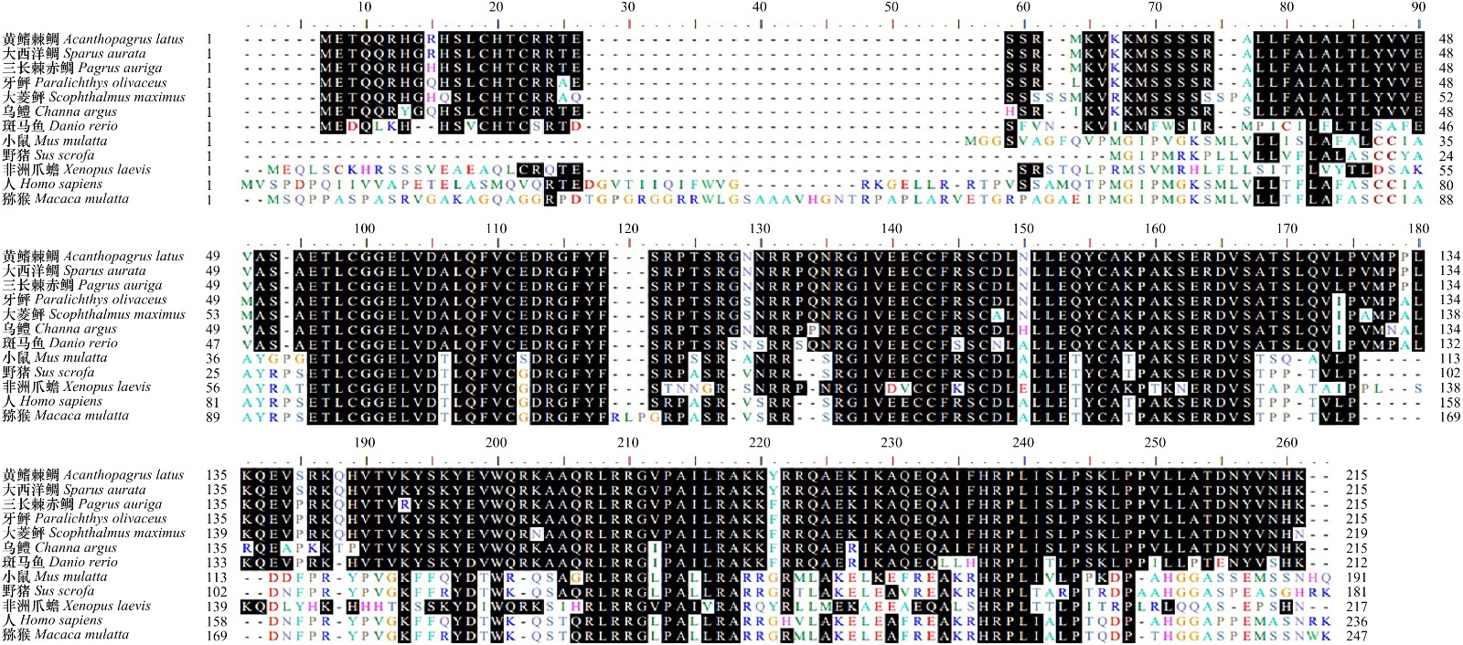
图 2 黄鳍棘鲷IGF-2基因氨基酸序列多重序列比对
不同颜色的字母代表不同的氨基酸;左右两边数字分别代表最左边与最右边的氨基酸在整个氨基酸序列中的位置
Fig. 2 Amino acid sequence multiple alignment of IGF-2 gene from Acanthopagrus latus
Different colored letters represent different amino acids; the left and right numbers represent the positions of the leftmost and rightmost amino acids in the amino acid sequence

图 3 黄鳍棘鲷和其他物种IGF-2基因氨基酸序列的系统进化树分析
Fig. 3 Phylogenetic analysis of IGF-2 gene amino acid sequence from Acanthopagrus latus and other species

图 4 黄鳍棘鲷的胚胎发育
a. 2细胞期;b. 4细胞期;c. 8细胞期;d. 16细胞期;e. 32细胞期;f. 64细胞期;g. 多细胞期;h. 桑葚胚期;i. 高囊胚期;j. 低囊胚期;k. 原肠前期;l. 原肠中期;m. 原肠晚期;n. 胚孔封闭期;o. 视囊形成期;p. 肌节出现期;q. 晶体形成期;r. 尾芽期;s. 尾芽游离期;t. 肌肉效应期;u. 心跳期;v. 将孵期;w. 破膜期
Fig. 4 Embryonic development of Acanthopagrus latus
a. 2-cell stage; b. 4-cell stage; c. 8-cell stage; d. 16-cell stage; e. 32-cell stage; f. 64-cell stage; g. multicellular stage; h. morula stage; i. high blastula stage; j. low blastula stage; k. early-gastrula stage; l. mid-gastrula stage; m. late-gastrula stage; n. closure of blastopore stage; o. eye vesicle formation stage; p. muscle burl stage; q. crystal formation stage; r. tail-bud stage; s. tail-bud free stage; t. muscular effect stage; u. heart-beating stage; v. pre-hatching stage; w. hatching stage
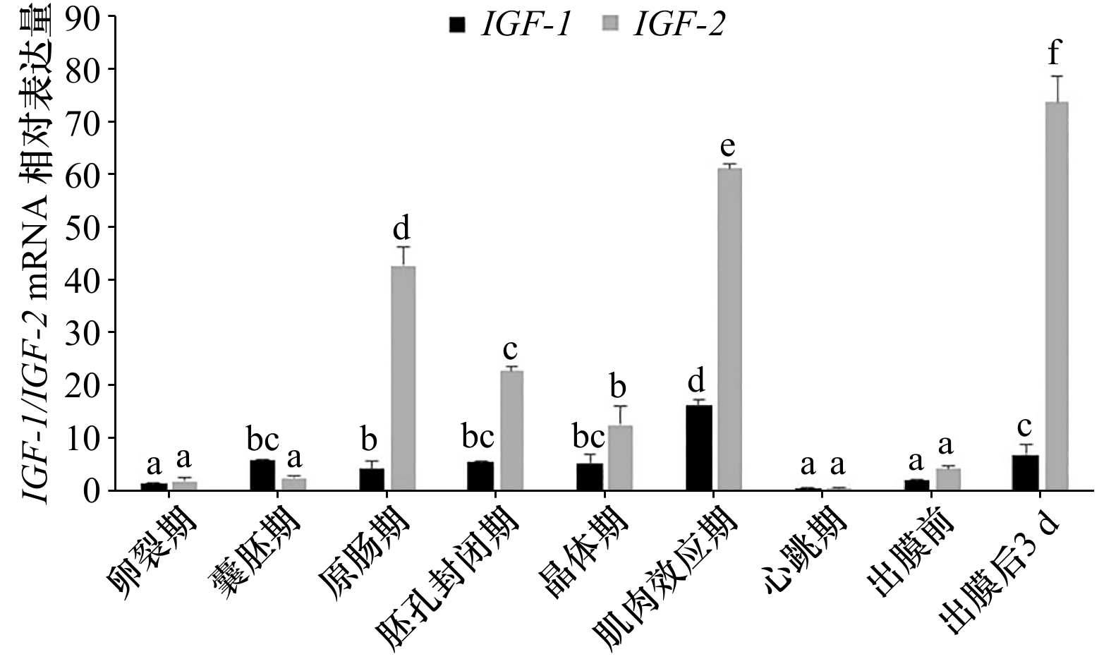
图 5 黄鳍棘鲷胚胎发育过程中IGF-1/IGF-2基因mRNA的相对表达量
标有不同字母的组间平均值差异显著(p<0.05)
Fig. 5 The relative expression of IGF-1/IGF-2 genes mRNA in different embryo development stages of Acanthopagrus latus
The mean values marked with different letters between groups are significantly striking(p<0.05)
表 1 本实验所用引物
Tab. 1 The primers used in the experiments
引物名称 序列(5′-3′) 用途 IGF-2-F TCATCTCAGCCGCACCAACT CDS序列克隆 IGF-2-R AAAAGGTGCTGGAACAGGAATC CDS序列克隆 5′-GSP1 GGCATCACGGGTAAGACCTGTA 5'末端序列克隆 5′-GSP2 GTGGCAAAGTGAGTGGCGTC 5'末端序列克隆 3′-GSP1 TCTGAACTCTTTCGCTCCCTCT 3'末端序列克隆 3′-GSP2 ATTAGATTCCTGTTCCAGCACCTT 3'末端序列克隆 M13F-47 CGCCACCCTTTTCCCAGTCACGAC 菌液检测 M13R-48 AGCGGATAACAATTTCACACAGGA 菌液检测 IGF-1-F1 TAGCCACACCCTCTCACTACTG 荧光定量 IGF-1-R1 AAGCCTCTCTCTCCACACACAA 荧光定量 IGF-2-F1 CCGTAGCTGTGACCTCAACC 荧光定量 IGF-2-R1 TCCTCTGCCACACCTCGTAT 荧光定量 β-actin-F ACCCAGATCATGTTCGAGACC 内参基因 β-actin-R ATGAGGTAGTCTGTGAGGTCG 内参基因  下载: 导出CSV
表 2 海水鱼类胚胎发育特征的比较
Tab. 2 Comparison of embryonic developmental characteristics of marine fish
物种 受精卵直径/
mm油球直径/
mm孵化水温/
°C胚胎发育
时间黄鳍棘鲷
(Acanthopagrus latus)0.900±0.050 0.250±0.020 26.5±0.5 25 h 30 min 条石鲷
(Oplegnathus fasciatus)[28]0.910±0.038 0.210±0.028 23.9±0.4 30 h 32 min 黑棘鲷
(Acanthopagrus schlegelii)[29]0.895±0.055 − 21.0±0.5 32 h 大西洋鲷
(Sparus aurata)[30]0.870±0.050 0.205±0.025 19±0.5 44 h 48 min 军曹鱼
(Rachycentron canadum)[31]1.245±0.065 0.325±0.027 27.0±0.5 26 h 30 min 鞍带石斑鱼
(Epinephelus lanceolatus)[32]0.820±0.030 0.230±0.030 29.0±0.5 18 h30 min 多纹钱蝶鱼
(Selenotoca multifasciata)[21]0.605±0.005 0.237±0. 013 27.0±1.0 18 h 30 min 黄姑鱼
(Nibea albiflora)[33]0.830±0.018 0.240±0.012 23.2±0.15 22 h 59 min
下载: 导出CSV
-
[1] Wood A W, Duan Cunming, Bern H A. Insulin-like growth factor signaling in fish[J]. International Review of Cytology, 2005, 243: 215−285. [2] Loir M, Le Gac F. Insulin-like growth factor-I and -II binding and action on DNA synthesis in rainbow trout spermatogonia and spermatocytes[J]. Biology of Reproduction, 1994, 51(6): 1154−1163. doi: 10.1095/biolreprod51.6.1154 [3] Zou Shuming, Kamei H, Modi Z, et al. Zebrafish IGF genes: gene duplication, conservation and divergence, and novel roles in midline and notochord development[J]. PLoS One, 2009, 4(9): e7026. doi: 10.1371/journal.pone.0007026 [4] Tse M C L, Vong Q P, Cheng C H K, et al. PCR-cloning and gene expression studies in common carp (Cyprinus carpio) insulin-like growth factor-II[J]. Biochimica et Biophysica Acta (BBA)-Gene Structure and Expression, 2002, 1575(1/3): 63−74. [5] Fukenstein B, Shemer R, Amuly R, et al. Nucleotide sequence of the promoter region of Sparus aurata insulin-like growth factor I gene and expression of IGF-I in eggs and embryos[J]. Molecular Marine Biology and Biotechnology, 1996, 5(1): 43−51. [6] White Y A R, Kyle J T, Wood A W. Targeted gene knockdown in zebrafish reveals distinct intraembryonic functions for insulin-like growth factor II signaling[J]. Endocrinology, 2009, 150(9): 4366−4375. doi: 10.1210/en.2009-0356 [7] 苏锦祥. 鱼类学与海水鱼类养殖[M]. 2版. 北京: 中国农业出版社, 2000: 160-220.Su Jinxiang. Ichthyology and Marine Fish Culture[M]. 2nd ed. Beijing: China Agriculture Press, 2000: 160−220. [8] 麦贤杰, 黄伟健, 叶富良, 等. 海水鱼类繁殖生物学和人工繁育[M]. 北京: 海洋出版社, 2005: 199-225.Mai Xianjie, Huang Weijian, Ye Fuliang, et al. Reproductive Biology and Artificial Breeding of Marine Fish[M]. Beijing: China Ocean Press, 2005: 199−225. [9] Wang S B, Lau K Y, Liu K M, et al. Reproductive characteristics of the hermaphroditic yellowfin seabream Acanthopagrus latus in the waters off western Taiwan[J]. Aquaculture Research, 2020, 51(12): 5015−5028. doi: 10.1111/are.14839 [10] 洪万树, 张其永, 郑建峰, 等. 港养黄鳍鲷性腺发育和性转变研究[J]. 台湾海峡, 1991, 10(3): 221−228.Hong Wanshu, Zhang Qiyong, Zheng Jianfeng, et al. Studies on gonadal development and sex inversion of yellowfin seabream (Acanthopagrus latus)[J]. Journal of Oceanography in Taiwan Strait, 1991, 10(3): 221−228. [11] 郑运通, 马荣和, 许波涛, 等. 黄鳍鲷人工繁殖与育苗技术的研究[J]. 海洋渔业, 1986(5): 205−208.Zheng Yuntong, Ma Ronghe, Xu Botao, et al. Study on artificial propagation and seedling technology of yellowfin bream (Acanthopagrus latus)[J]. Marine Fisheries, 1986(5): 205−208. [12] 郑运通, 马荣和, 许波涛, 等. 黄鳍鲷的胚胎和仔稚幼鱼的形态发育观察[J]. 水产科技情报, 1986(4): 1−3.Zheng Yuntong, Ma Ronghe, Xu Botao, et al. Morphological development of embryos and larvae of yellowfin bream (Acanthopagrus latus)[J]. Fisheries Science and Technology Information, 1986(4): 1−3. [13] Leu M Y, Chou Y H. Induced spawning and larval rearing of captive yellowfin porgy, Acanthopagrus latus (Houttuyn)[J]. Aquaculture, 1996, 143(2): 155−166. doi: 10.1016/0044-8486(96)01272-0 [14] Li Shizhu, Lin Genmei, Fang Wenyu, et al. Gonadal transcriptome analysis of sex-related genes in the protandrous yellowfin seabream (Acanthopagrus latus)[J]. Frontiers in Genetics, 2020, 11: 709. doi: 10.3389/fgene.2020.00709 [15] Zhou Ying, Liu Haiyang, Wang Xinhua, et al. QTL fine mapping for sex determination region in bighead carp (Hypophthalmichthys nobilis) and comparison with silver carp (Hypophthalmichthys molitrix)[J]. Marine Biotechnology, 2020, 22(1): 41−53. doi: 10.1007/s10126-019-09929-3 [16] Zhu Kecheng, Zhang Nan, Liu Baosuo, et al. A chromosome-level genome assembly of the yellowfin seabream (Acanthopagrus latus; Hottuyn, 1782) provides insights into its osmoregulation and sex reversal[J]. Genomics, 2021, 113(4): 1617−1627. doi: 10.1016/j.ygeno.2021.04.017 [17] 石和荣, 张为民, 刘晓春, 等. 半胱胺盐酸盐和 LHRH-A 对黄鳍鲷生长激素分泌的影响[J]. 海洋学报, 2005, 27(3): 147−153.Shi Herong, Zhang Weimin, Liu Xiaochun, et al. Effects of cysteamine hydrochloride and luteinizing hormone-releasing hormone analog on growth hormone secretion in yellowfin porgy[J]. Haiyang Xuebao, 2005, 27(3): 147−153. [18] 石和荣, 张勇, 张为民, 等. 半胱胺盐酸盐和LHRH-A对黄鳍鲷IGF-I基因表达和生长的影响[J]. 动物学报, 2005, 51(1): 108−116. doi: 10.3969/j.issn.1674-5507.2005.01.016Shi Herong, Zhang Yong, Zhang Weimin, et al. Effect of cysteamine hydrochloride and luteininzing hormone-releasing hormone analog on the growth and the expression of IGF-I mRNA in the yellowfin porgy Sparus latus[J]. Acta Zoologica Sinica, 2005, 51(1): 108−116. doi: 10.3969/j.issn.1674-5507.2005.01.016 [19] 张殿昌, 江世贵. 黄鳍鲷生长激素cDNA的分子克隆和序列分析[J]. 湛江海洋大学学报, 2002, 22(4): 62−65.Zhan Dianchang, Jiang Shigui. Molecular cloning and sequence analysis of growth hormone cDNA from Sparus latus[J]. Journal of Zhanjiang Ocean University, 2002, 22(4): 62−65. [20] 马细兰, 冷婷婷, 刘启智, 等. 黄鳍鲷(Sparus latus)两种生长激素受体的cDNA克隆及组织表达分析[J]. 海洋与湖沼, 2011, 42(6): 830−838. doi: 10.11693/hyhz201106013013Ma Xilan, Leng Tingting, Liu Qizhi, et al. cDNAs cloning and tissues expression of two growth hormone receptors in yellowfin bream Sparus latus[J]. Oceanologia et Limnologia Sinica, 2011, 42(6): 830−838. doi: 10.11693/hyhz201106013013 [21] 刘鉴毅, 李琪, 孙艳秋, 等. 多纹钱蝶鱼胚胎发育及胚后发育观察[J]. 中国水产科学, 2021, 28(8): 978−987.Liu Jianyi, Li Qi, Sun Yanqiu, et al. Embryonic and post-embryonic development of Selenotoca multifasciata[J]. Journal of Fishery Sciences of China, 2021, 28(8): 978−987. [22] 杨慧荣, 王庆, 李水生, 等. 类胰岛素生长因子(IGFs)在斜带石斑鱼胚胎及卵巢的表达[J]. 中山大学学报(自然科学版), 2019, 58(5): 94−103.Yang Huirong, Wang Qing, Li Shuisheng, et al. Expression analysis of insulin-like growth factors’ (IGFs) in embryo and oocyte of Epinephelus coioides[J]. Acta Scientiarum Naturalium Universitatis Sunyatseni, 2019, 58(5): 94−103. [23] Aslan O, Hamill R M, Davey G, et al. Variation in the IGF2 gene promoter region is associated with intramuscular fat content in porcine skeletal muscle[J]. Molecular Biology Reports, 2012, 39(4): 4101−4110. doi: 10.1007/s11033-011-1192-5 [24] Yuan Yongming, Hong Yunhan. Medaka insulin-like growth factor-2 supports self-renewal of the embryonic stem cell line and blastomeres in vitro[J]. Scientific Reports, 2017, 7(1): 78. doi: 10.1038/s41598-017-00094-y [25] 林权卓, 沈卓坤, 杨宪宽, 等. 双棘黄姑鱼IGF2基因克隆及其在卵巢发育中的作用研究[J]. 广东农业科学, 2015, 42(3): 119−124, 130. doi: 10.3969/j.issn.1004-874X.2015.03.026Lin Quanzhuo, Shen Zhuokun, Yang Xiankuan, et al. IGF2 gene cloning and its function during the development of ovarian cycle in Protonibea diacantus[J]. Guangdong Agricultural Sciences, 2015, 42(3): 119−124, 130. doi: 10.3969/j.issn.1004-874X.2015.03.026 [26] 王丁科, 阎萍, 梁春年, 等. 胰岛素样生长因子2研究进展[J]. 动物医学进展, 2008, 29(7): 67−70. doi: 10.3969/j.issn.1007-5038.2008.07.017Wang Dingke, Yan Ping, Liang Chunnian, et al. Progress on insulin-like growth factor 2[J]. Progress in Veterinary Medicine, 2008, 29(7): 67−70. doi: 10.3969/j.issn.1007-5038.2008.07.017 [27] 陈军平, 沈方方, 武慧慧, 等. 我国鱼类胚胎发育研究进展[J]. 江苏农业科学, 2021, 49(17): 45−52.Chen Junping, Shen Fangfang, Wu Huihui, et al. Research progress of China’s fish embryonic development[J]. Jiangsu Agricultural Sciences, 2021, 49(17): 45−52. [28] 辛俭, 薛利建, 毛国民, 等. 条石鲷的胚胎发育观察[J]. 浙江海洋学院学报(自然科学版), 2005, 24(1): 31−36.Xin Jian, Xue Lijian, Mao Guomin, et al. Study on the embryonic development of Oplegnathidae fasciatus[J]. Journal of Zhejiang Ocean University (Natural Science), 2005, 24(1): 31−36. [29] 官曙光, 刘洪军, 李祥东, 等. 黑棘鲷胚胎发育过程及特殊结构观察[J]. 海洋科学, 2011, 35(9): 68−72.Guan Shuguang, Liu Hongjun, Li Xiangdong, et al. Observation of embryonic development of Acanthopagrus schlegelii[J]. Marine Sciences, 2011, 35(9): 68−72. [30] 王彦怀, 陶秉春, 梁伟光, 等. 金头鲷胚胎发育的初步观察[J]. 海洋水产研究, 2006, 27(6): 14−18.Wang Yanhuai, Tao Bingchun, Liang Weiguang, et al. Preliminary studies on embryo development of Sparus aurata[J]. Marine Fisheries Research, 2006, 27(6): 14−18. [31] 邝杰华, 陈刚, 马骞, 等. 军曹鱼的胚胎发育及仔稚鱼形态观察[J]. 水产学报, 2021, 45(11): 1814−1824.Kuang Jiehua, Chen Gang, Ma Qian, et al. Embryonic development and morphological characteristics of larvae and juveniles of cobia (Rachycentron canadum)[J]. Journal of Fisheries of China, 2021, 45(11): 1814−1824. [32] 周玲, 翁文明, 李金亮, 等. 鞍带石斑鱼胚胎发育及仔鱼形态发育、饵料转变的观察研究[J]. 中国农学通报, 2010, 26(1): 293−302.Zhou Ling, Weng Wenming, Li Jinliang, et al. Studies on embryonic development, morphological development and feed changeover of Epinephelus lanceolatus larva[J]. Chinese Agricultural Science Bulletin, 2010, 26(1): 293−302. [33] 黄贤克, 单乐州, 闫茂仓, 等. 黄姑鱼胚胎发育及其与温度和盐度的关系[J]. 海洋科学, 2017, 41(7): 44−50. doi: 10.11759/hykx20160920003Huang Xianke, Shan Lezhou, Yan Maocang, et al. Embryonic development of Nibea albiflora and the effects of temperature and salinity on embryogenesis[J]. Marine Sciences, 2017, 41(7): 44−50. doi: 10.11759/hykx20160920003 [34] Yang S G, Ji S C, Lim S G, et al. Management of sexual maturation and natural spawning of captive-reared yellowtail kingfish, Seriola lalandi, in an indoor rearing tank[J]. Development & Reproduction, 2016, 20(2): 141−147. [35] 张克伟, 陈华谱, 江东能, 等. 金钱鱼IGF-1和IGF-2的克隆及其在胚胎发育过程的表达[J]. 广东海洋大学学报, 2018, 38(2): 7−14. doi: 10.3969/j.issn.1673-9159.2018.02.002Zhang Kewei, Chen Huapu, Jiang Dongneng, et al. Insulin-like growth factors 1 and 2 in spotted scat (Scatophagus argus): molecular cloning and differential expression during embryonic development[J]. Journal of Guangdong Ocean University, 2018, 38(2): 7−14. doi: 10.3969/j.issn.1673-9159.2018.02.002 [36] Perrot V, Moiseeva E B, Gozes Y, et al. Ontogeny of the insulin-like growth factor system (IGF-I, IGF-II, and IGF-1R) in gilthead seabream (Sparus aurata): expression and cellular localization[J]. General and Comparative Endocrinology, 1999, 116(3): 445−460. doi: 10.1006/gcen.1999.7337 [37] Greene M W, Chen T T. Quantitation of IGF-I, IGF-II, and multiple insulin receptor family member messenger RNAs during embryonic development in rainbow trout[J]. Molecular Reproduction and Development, 1999, 54(4): 348−361. doi: 10.1002/(SICI)1098-2795(199912)54:4<348::AID-MRD5>3.0.CO;2-N -

计量
- 文章访问数: 682
- HTML全文浏览量: 395
- PDF下载量: 67
- 被引次数: 0
